Home

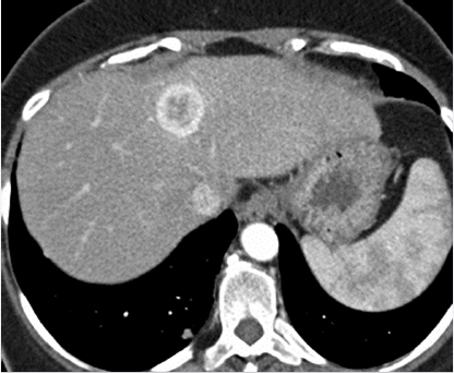
afternoon Quote Lima ring enhancing lesion liver Angry wall Relationship

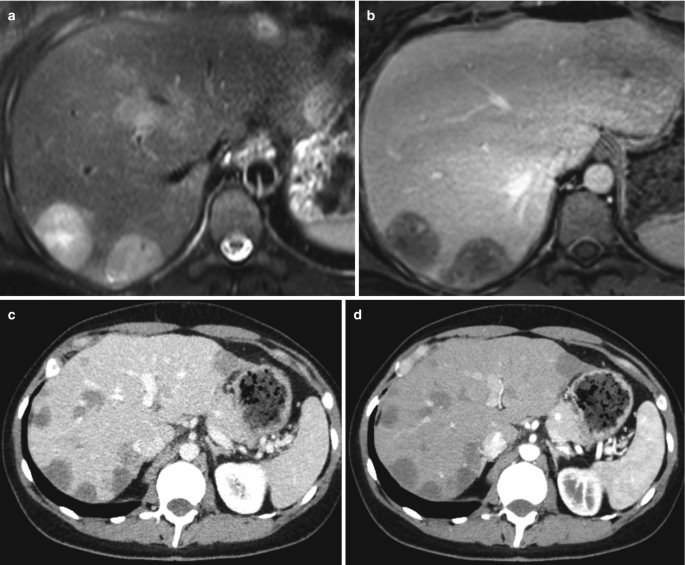
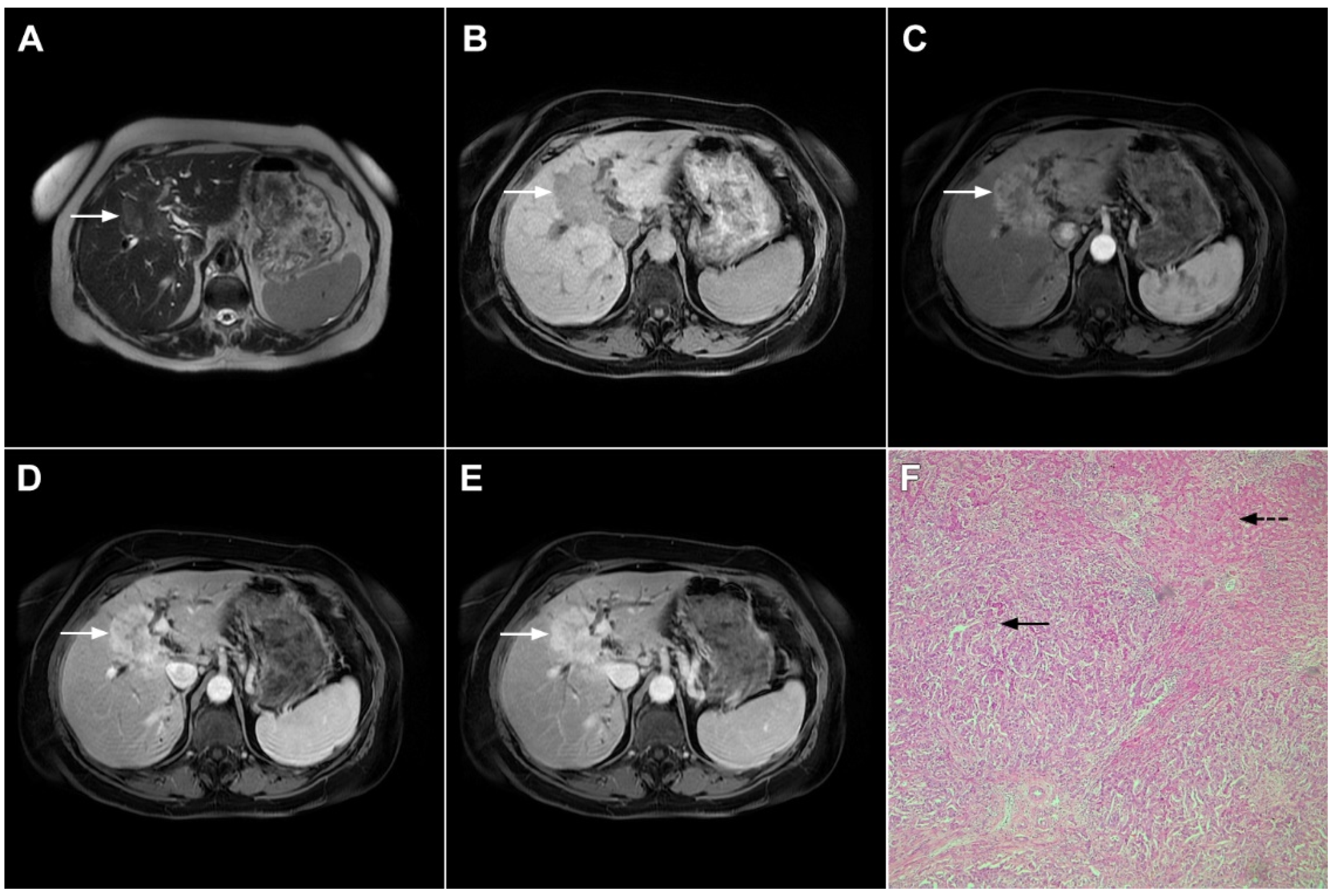
Current Oncology | Free Full-Text | Imaging Spectrum of Intrahepatic  Mass-Forming Cholangiocarcinoma and Its Mimickers: How to Differentiate  Them Using MRI
Current Oncology | Free Full-Text | Imaging Spectrum of Intrahepatic Mass-Forming Cholangiocarcinoma and Its Mimickers: How to Differentiate Them Using MRI

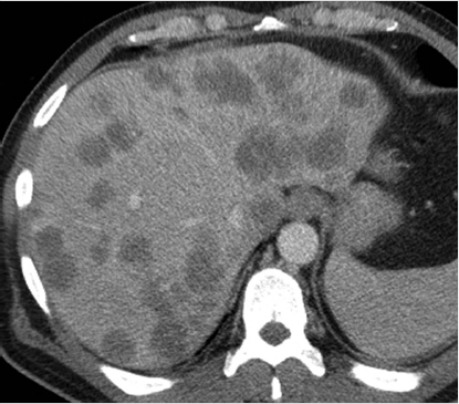
CT scan of abdomen and pelvis with contrast showing multiple hepatic... |  Download Scientific Diagram
CT scan of abdomen and pelvis with contrast showing multiple hepatic... | Download Scientific Diagram

Spectrum of liver lesions hyperintense on hepatobiliary phase: an approach  by clinical setting | Insights into Imaging | Full Text
Spectrum of liver lesions hyperintense on hepatobiliary phase: an approach by clinical setting | Insights into Imaging | Full Text
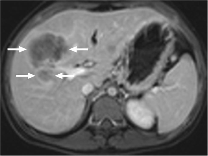
Ring-enhancing hepatocellular carcinoma, indicative of a more... | Download  Scientific Diagram
Ring-enhancing hepatocellular carcinoma, indicative of a more... | Download Scientific Diagram

Hypervascular Liver Lesions - ScienceDirect

14 Imaging of Liver Tumors | Radiology Key

Hypervascular Liver Lesions - ScienceDirect

Non-neoplastic hepatopancreatobiliary lesions simulating malignancy: can we  differentiate? | Insights into Imaging | Full Text
Non-neoplastic hepatopancreatobiliary lesions simulating malignancy: can we differentiate? | Insights into Imaging | Full Text

Focal Liver Lesions | SpringerLink

EPOS™
EPOS™

Role of MDCT in Detection and Characterisation of Focal Liver Lesions

Ring enhancement (A) and subsequent ring oil deposition (B) on a CRLM... |  Download Scientific Diagram
Ring enhancement (A) and subsequent ring oil deposition (B) on a CRLM... | Download Scientific Diagram

Imaging results. (a) Computed tomography shows a ring-enhancing 28-mm... |  Download Scientific Diagram
Imaging results. (a) Computed tomography shows a ring-enhancing 28-mm... | Download Scientific Diagram

CT scan showing two of the five ring-enhancing nodular lesions. | Download  Scientific Diagram
CT scan showing two of the five ring-enhancing nodular lesions. | Download Scientific Diagram

The Radiology Assistant : Characterisation of liver masses

Snehal Lapsia on X: "74 yrs. Normal background liver. High T1 signal, ring  enhancing lesion ?Haemorrhagic or melanoma metastasis Confirmed melanoma  met #FOAMrad #FOAMed #MedEd #MedTwitter #liver #HBP #surgery #radiology  #dermatology #anatomy #
Snehal Lapsia on X: "74 yrs. Normal background liver. High T1 signal, ring enhancing lesion ?Haemorrhagic or melanoma metastasis Confirmed melanoma met #FOAMrad #FOAMed #MedEd #MedTwitter #liver #HBP #surgery #radiology #dermatology #anatomy #
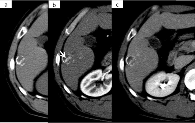
The Radiology Assistant : LI-RADS

Hyperechoic liver lesions | Radiology Reference Article | Radiopaedia.org

Hepatocellular carcinoma with ring calcification mimicking hydatid disease:  a case report | Surgical Case Reports | Full Text
Hepatocellular carcinoma with ring calcification mimicking hydatid disease: a case report | Surgical Case Reports | Full Text

The Radiology Assistant : Characterisation of liver masses

The Radiology Assistant : LI-RADS

Focal Liver Lesions | SpringerLink

A case of macronodular hepatic tuberculosis in a young African male |  Eurorad
A case of macronodular hepatic tuberculosis in a young African male | Eurorad

14 Imaging of Liver Tumors | Radiology Key

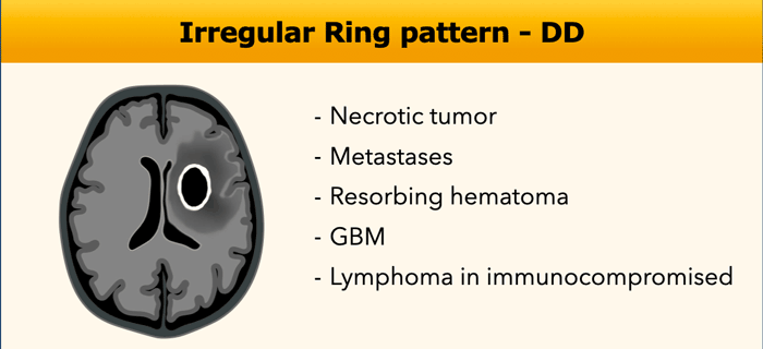
The Radiology Assistant : Enhancement Patterns in CNS disease